La tecnología Bexel se desarrolló por primera vez a través de la colaboración entre varios científicos rusos y coreanos en 1997.
Una conocida empresa coreana establecida llamada STC (conocida por su desarrollo masivo de baterías y maquinillas de afeitar Gillette) se involucró en una empresa conjunta con el gobierno coreano que luego estableció rigurosamente el protocolo de investigación y desarrollo iridológico Bexel Irna con sede en la Universidad de la Amistad de los Pueblos. de Rusia.
Gran parte de la base de esta tecnología se basó en el iridólogo más prestigioso de Rusia, el Dr. Velchover.
Lo más especial de la tecnología Bexel Irina es que la tabla topográfica utilizada en el software se coloca con precisión en cada ojo único, por lo tanto, cada tabla es única para cada conjunto de ojos.
El sistema Bexel fue el único sistema de análisis iridológico que alguna vez logró con éxito el estado aprobado por la FDA como dispositivo médico clase 3 en Corea, luego de los exitosos estudios clínicos certificados por el gobierno coreano realizados en la Universidad AJU.
Puede leer los resultados del estudio clínico del hospital Bexel aquí: Estudio clínico Bexel Irina - Universidad de Aju
Tras el colapso de la economía asiática en 1998, Bexel Irina nunca llegó al mercado norteamericano o europeo. Sin embargo, algunos sistemas se vendieron a algunos hospitales ubicados a lo largo del borde asiático.
¿Por qué la tecnología Bexel Irina sigue siendo útil para la iridología?
La tecnología Bexel Irina se basa en la bio-métrica del ojo y ofrece parámetros matemáticos muy valiosos para la pupila y el collarín. Estos parámetros matemáticos son muy importantes para verificar los cambios en el sistema nervioso autónomo que pueden usarse para estudios clínicos y determinar los cambios ACTUALES en el ojo humano, incluida la mejora de la salud del paciente o una patología en curso. ¡Los estudios clínicos requieren evidencia científica sólida y Bexel Irina puede entregar esa información científica!
En el pasado, muchos iridólogos cometieron el error de determinar la patología de la salud actual basándose en varios signos en el iris, incluidas las lagunas. Como entendemos ahora, las lagunas son genotípicas, no cambian de densidad, por lo que no siempre representan patología de la enfermedad activa. Sin embargo, cuando hay signos asociados con lagunas como las deformaciones del collarete o la pupila, el iridólogo puede establecer una patología activa asociada con la debilidad genética (lagunas). En otras palabras, si su paciente logra con éxito un protocolo de salud con una mejora notable, no verá "líneas de cicatrización" en las lagunas, pero debería ver cambios positivos en los parámetros pupilares. Y los cambios en la pupila pueden probarse científicamente con los datos de parámetros matemáticos de Bexel Irina.
Para el iridólogo, es muy difícil determinar los diversos grados de deformaciones, descentraciones y multiformaciones de la pupila. Por ejemplo, la planitud pupilar, no menos de una sexta parte de la circunferencia de la pupila, se considera significativa para mostrar patología activa. ¡Para un humano detectar este grado de planitud sería una tarea muy difícil!
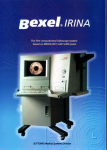
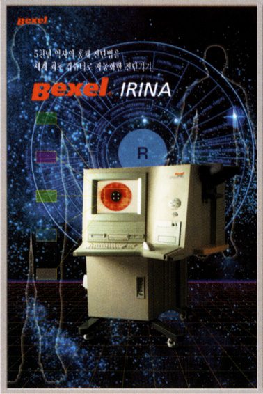
STC (Suttong) invirtió más de 10 millones de dólares en el desarrollo de Bexel Irina. El sistema Bexel que se muestra en las imágenes adjuntas se ofreció a los hospitales a 35K USD. Sería una pena que esta tecnología fuera olvidada y por qué Americana De Salud ofrecerá esta increíble tecnología a sus miembros a través de nuestra plataforma.
Solicite informacion: asenat.cursos@yahoo.com/ Whatsapp al final esta el enlace.
What is the Bexel Irina Technology? The Bexel Technology was first developed through collaboration between several Russian and Korean scientists in 1997. A well known established Korean company named STC became involved in a joint venture with the Korean Government who then rigorously established the Bexel Irna iridological research and development protocal based at the The Peoples’ Friendship University of Russia. Much of the basis of this technology was founded upon Russia’s most prestigious iridologist, Dr. Velchover. The Bexel system was the only iridological analysis system that ever successfully achieved FDA approved status as medical device class 3 in Korea, following the highly successful Korean government certified clinical studies held at AJU University. You can read the results of the Bexel Clinical Hospital Study here:
Why is the Bexel Irina Technology still useful to Iridology?
The Bexel Irina technology is based on eye bio-metrics and offers very valuable mathematical parameters of the pupil and collarette. These mathematical parameters are very important in verifying changes in the autonomic nervous system that can be used for clinical studies and determining ACTUAL changes in the human eye, including patient health improvement or an ongoing pathology. Clinical studies require strong scientific evidence and the Bexel Irina can deliver that scientific data!
Correcting Past Mistakes
Previously many individuals in the iridological sciences made the mistake of determining current health pathology based upon various signs in the iris including lacunae. As we now understand, lacunae are genotypic, do not change in density, thus do not always represent active disease pathology. However, when there are associated signs with lacunae such as collarette or pupil deformations, we may then establish an active pathology associated with genetic weakness (lacunae). In other words, if your patient successfully achieves a health protocol with notable improvement, you will not see ‘healing lines’ in lacunae, but you should see positive changes in the pupillary parameters. And these changes in pupil can be scientifically proven with the Bexel Irina mathematical parameter data.
Difficulty in Pupil and Collarette Analysis for Humans
For most individuals, it can be very difficult to determine the several degrees of pupil deformations, decentrations and multiformaties. For example, pupillary flatness, not less than one sixth of pupil circumference, is considered to be significant in showing active pathology. For a human to detect this degree of flatness would be a very difficult task!
Over 10 Million USD Investment
STC (Suttong) invested countless man hours including over 10 million USD into the Bexel Irina development. The Bexel system displayed in the displayed images on this page was offered to hospitals throughout Asia for 35K USD. Following the collapse of Asian economics back in 1998, the Bexel Irina never was available to the North American or European market. However, a few systems were sold to some hospitals located along the Asian rim. It would be a shame if this technology was forgotten and why we will continue to offer this amazing technology to the public and professional health clinics!
Korean Hospital Study using Bexel Irina Iridological Analysis System
Methods:
352 patients with different diseases (mostly of the hepatic-bilary system) were examined under hospital conditions. Many of them were relatives and representatives of different generations of the family. Patients were 14 to 83 years old and there were 126 men (35.8%) and 226 women (64.2%).
Iridology examinations were carried out with the BEXEL IRINA device. The image of the iris was placed into the computer memory and displayed on screen with the help of the optical system with illumination camera, and special input units of the optical equipment connected to the computer. The iris images are converted into graphic files. Iridologists can see movements of the iris structures in real time mode or they can fix the selected images. The advanced iridogiagnostic software of the system provides computer processing of the iris picture, automatic determination of the main iris structure and automatic measurement of the geometric parameters, the area of the irises, the area, form, the position of the pupils, width and form of the pupillary margin, and the form of the collarette. The first part of the iris evaluation is based on the processing of these parameters, is also done automatically.
The second stage of the diagnosis is based on the mapping of the iris according to the selected chart. Designed especially and the newest iridological chart, including the achievements of modern iridology, was developed by Dr. Mararchuk. Superposition of the chart on the iris of the person is also done automatically with the corresponding adjustments to the borders of the iris structures of the person. Hardware and software are adapted to the dark pigment-saturated type of irises of Asian people.
The conclusions made by iridology examinations were compared with the data of other medical diagnostic methods carried out both before and after the examination (biochemical X-ray, endoscope, angiographic radioisotope and intra-operative examinations, tetrapolar rheography, ultrasonic study, computer tomography, mammography, etc.).
Results of Study
The results of 352 patients were analyzed. There were 170 patients with chronic calculous cholecystis (15 men and 145 women). Forty-nine patients with chronic non-calculous cholecystitis (12 men and 37 women), 66 patients with hepatitis and cirrhosis (44 men and 22 women). Forty-two patients with portal hypertension (26 men and 16 women). In patients with chronic calculous cholecystitis, the deformation of the pupils was marked in 39% of the patients and was projected at the right side of the hepatic-biliary system in 22% and the left side in 17%. According to the majority of the iridological charts, the projection of the gall bladder is defined within 7.30 – 8.00 of the ciliary belt of the right iris. In the area of the iris, we revealed different changes in 132 patients, which was 77.6%. The basic manifestations of calculous cholecystitis on the iris of the right eye were pigment spots with different shapes, lacunae etc.
Generally, the pronunciation of the iris signs depended upon the duration and severity of the pathological process. The outlined stroma, the areas of clarification, the rare pigment enclosures and small lacunae took place in 74.6% of the patients in which pathology was occasionally revealed. These patients were asymptomatic in their study of disease, had a duration of disease of less than two years, and had no hereditary parallels for disease. In the case of diseases with duration of more than three years, frequent exacerbations and distinct indication to the hereditary character of the process changes were present in the hepatic-bilary area of the iris in 92.6% of the cases. Pigment spots occurred in 69% of the cases and deep lacunae, with thick walls in combination with pigment spots occurred in 18% of the cases.
In 22% of the patients with concrements in the gall bladder, there were no iridological signs on the irises.
Similar data were obtained for patients with chronic non-calculous cholecystitis (49%). The local changes in the gall bladder sector of the right iris were revealed in 77.6% of the patients. Most often, (66% of the patients) an iridological sign of non-calculous cholecystitis is shown by the local whitish thickening of the iris stroma in this sector zone or a non-linear clarification of the stroma. Comparing the inheritance of cholecystitis in the different generations of families, we have come to the conclusion that this pathology is associated with sex and is inherited mostly by females.
The most frequently accompanying disease of chronic cholecystitis are: chronic pancreatitis, cardiac ischemic disease, gastro-duodenal pathology and kidney diseases. The most common iridological signs in people with portal hypertension (42) were pigmentation of the autonomous wreath (74%), internal heterochromy (31%), changes in the right iris in the 7.30-8.10 sector (87%), and drawing-in of the autonomous wreath in the liver area of the right iris (76%).
In many of our cases, pathological iris signs in the liver projections were found in people having no clinical symptoms of any disease. Detailed laboratory examinations of these people revealed initial changes in the hepatic-biliary system in 63% of these people. Such signs can be considered as the markings of hereditary risk of hepatic-biliary pathology which can develop in the future. The correct prophylactic measures such as diet etc. can help prevent disease.
Golden Standards: The Suttong Clinical Iridological Project
These studies were conducted at AJU University General Hospital located in Seoul, South Korea. They ultimately succeeded to obtain an impressive government certification in 1998. These were the outcomes.
Changes on the ins in the malignant pathology of the breast can be registered three to five years before the system complex really starts to occur.
In some types of lung cancer, as early as two to three years before symptoms appear the iris will change.
In people at risk for cardio-spastic reactions, iris signs are evident 0.5 to two years before symptoms appear.
In children the functional weakness of the pancreas, gall bladder, and the risk of broncho-spastic reactions are well verified in the iris. Moreover, a 10-year master prospective study was held in Korea complete with these outcomes: all children and teenagers detected via the iris examination as belonging to the risk groups systematically underwent preventive measures (diet, special gymnastics, etc.) They showed a significant percentage of decrease in manifestation of their genetic pathology when compared with the control data.
The study revealed the opportunity to analyze the state of an organism from the rare viewpoint of its integrity.
The study greatly promoted such features as completeness and reliability of the iris diagnosis.
As Joseph Deck himself once stated, irido-diagnostics are not limited by the thick clinical frameworks of one organ, but makes room for finding all the places of lowest resistance in the organism.
By revealing primary vs. secondary changes in the body, iridological examination is actively searching for the main primary component that violates the process of vital activity and not for the secondary actors in the pathological process.